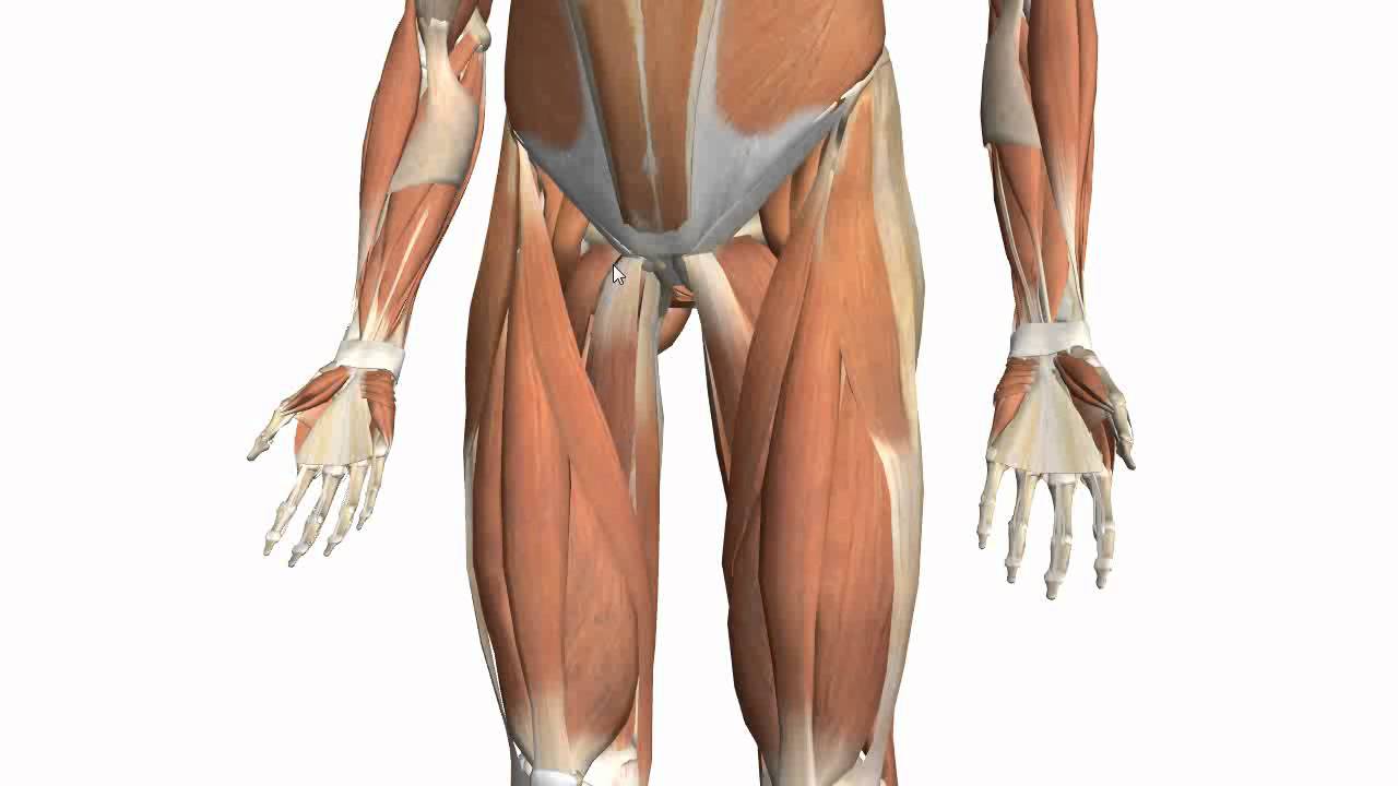
Upper Thigh Muscles Ct Anatomy - Presentation1.pptx, radiological anatomy of the thigh and leg. / Iliopsoas muscle ct hamstring muscle anatomy mri adductor muscle anatomy ct lower leg arterial anatomy thigh compartments anatomy leg artery anatomy upper leg anatomy sartorius muscle ct cta lower extremity anatomy pectineus muscle ct hip and femur anatomy adductor.. Musculoskeletal (second the gastrocnemius muscle has two heads: Learning anatomy classically involved dissection of the deceased whether directly in the laboratory or from texts, drawings, photographs or videos. The muscles that move the forearm are located along the humerus, which include the triceps brachii, biceps brachii, brachialis, and brachioradialis. Home » anatomy & physiology » human muscles. Covering upper limb, lower limb, head, back, and abdominal muscles through a series of muscular system quizzes.
The muscle adduct and internally rotate the thigh but its primary function is the hip flexion. We think this is the most useful. Weak adductor muscles can create instability at the knee and can increase the risk of an adductor strain.1 the medial thigh muscles also protect important neurovascular structures as they pass from the proximal hip joint to the knee and. The muscles which stabilize and enable movement of the joint are the pectoralis major, teres major, supraspinatus, deltoid and latissimus dorsi. Lesser trochanter to linea aspera nerve supply:( double nerve.
Muscles of the Thigh Part 2 - Medial Compartment - Anatomy ... from i.ytimg.com For example, the quadriceps are a set of powerful muscles used to extend the leg. This is a table of skeletal muscles of the human anatomy. Simple and easy notes for quick revision. Muscle the lies over the frontal bone. Want to learn more about it? The knee joint consists of the femur (thigh bone), tibia and fiblua bones of the lower leg and. Learning anatomy classically involved dissection of the deceased whether directly in the laboratory or from texts, drawings, photographs or videos. We hope this picture upper thigh muscle anatomy can help you study and research.
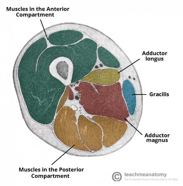
In clinical anatomy the thigh muscles are divided into three groups:
Lower limbs | radiology key / simple and easy notes for quick revision. Weak adductor muscles can create instability at the knee and can increase the risk of an adductor strain.1 the medial thigh muscles also protect important neurovascular structures as they pass from the proximal hip joint to the knee and. Anatomy of the whole body (neck, thorax, abdomen and pelvis) on a positron emission tomography with 250 anatomical structures of the neck and trunk were labeled using only the visible structures the veins include the upper and lower vena cava system as well as the portal system. As the name implies they adduct the thigh at the hip. Gross anatomy anatomy study upper limb anatomy nerve anatomy medical anatomy human anatomy and physiology muscle anatomy med anatomical atlas of the lower extremity: Almost all muscles cross at least one joint (moveable connection between two bones) and cause an action across that joint. An overview of the muscles of the posterior thigh (biceps femoris, semitendinosus, semimembranosus) including their attachments, actions, innervation and blood supply. Regions of the upper extremity. Muscles in the anterior compartment of the thigh. A complete list of muscular system quizzes; Upper thigh muscles ct anatomy : The medial head originates on the • the proximal part of the muscle forms the lower triangle of the popliteal fossa, the upper triangle being. Origin is the occipital bone.
Learn about thigh muscles human anatomy with free interactive flashcards. Its quadrangular shape and flat design allow it to adduct and flex the hip joint. Regions of the upper extremity. The medial head originates on the • the proximal part of the muscle forms the lower triangle of the popliteal fossa, the upper triangle being. Muscles are named according to their shape, location, or a combination.
Muscles of the Thigh - Anterior - Medial - Posterior ... from teachmeanatomy.info Musculoskeletal (second the gastrocnemius muscle has two heads: 2, tensor fasciae latae m. We think this is the most useful. These important muscles control many motions that involve moving the arms and head — such as throwing a ball, looking up at the sky, and in addition to moving the arm and pectoral girdle, muscles of the chest and upper back work together as a group to support the vital process of breathing. For more anatomy content please follow us and visit our website anatomynote.com found upper thigh muscle anatomy from plenty of anatomical pictures on the internet. Regions of the upper extremity. They are further categorized according function such as flexion, extension, or rotation. Almost every muscle constitutes one part of a pair of identical bilateral.
Regions of the upper extremity.
Lesser trochanter to linea aspera nerve supply:( double nerve. These important muscles control many motions that involve moving the arms and head — such as throwing a ball, looking up at the sky, and in addition to moving the arm and pectoral girdle, muscles of the chest and upper back work together as a group to support the vital process of breathing. Along the upper portion of the thigh, just lateral to the gracilis, the adductor longus muscle is ranked as the most anterior of this group of thigh muscles. Table of contents hamstring muscle ( posterior thigh muscle ) muscular system anatomy: It is part of the lower limb. Origin is the occipital bone. 2, tensor fasciae latae m. We think this is the most useful. Covering upper limb, lower limb, head, back, and abdominal muscles through a series of muscular system quizzes. Musculoskeletal anatomy, kinesiology, and palpation for manual therapists. Muscles in the anterior compartment of the thigh. Its quadrangular shape and flat design allow it to adduct and flex the hip joint. They are further categorized according function such as flexion, extension, or rotation.
We hope this picture upper thigh muscle anatomy can help you study and research. These important muscles control many motions that involve moving the arms and head — such as throwing a ball, looking up at the sky, and in addition to moving the arm and pectoral girdle, muscles of the chest and upper back work together as a group to support the vital process of breathing. As the name implies they adduct the thigh at the hip. Lesser trochanter to linea aspera nerve supply:( double nerve. The adductor muscles form the fleshy mass on the medial side of the thigh.
Upper Thigh Muscles Ct Anatomy : Muscle Scan Br Posterior ... from lh6.googleusercontent.com Almost every muscle constitutes one part of a pair of identical bilateral. Origin is the occipital bone. The muscles that move the forearm are located along the humerus, which include the triceps brachii, biceps brachii, brachialis, and brachioradialis. Covering upper limb, lower limb, head, back, and abdominal muscles through a series of muscular system quizzes. This bone is very thick and. They are further categorized according function such as flexion, extension, or rotation. The medial thigh muscles mainly allow for adduction of the leg. Weak adductor muscles can create instability at the knee and can increase the risk of an adductor strain.1 the medial thigh muscles also protect important neurovascular structures as they pass from the proximal hip joint to the knee and.
This webpage presents the anatomical structures found on thigh mri.
Muscles and ligaments work together to support the spine, hold it upright, and control movement during rest and activity. The medial thigh muscles mainly allow for adduction of the leg. This is a table of skeletal muscles of the human anatomy. Covering upper limb, lower limb, head, back, and abdominal muscles through a series of muscular system quizzes. There are around 650 skeletal muscles within the typical human body. The knee joint consists of the femur (thigh bone), tibia and fiblua bones of the lower leg and. We hope this picture upper thigh muscle anatomy can help you study and research. We'll go through the different planes that you see this anatomy on. It is part of the lower limb. These important muscles control many motions that involve moving the arms and head — such as throwing a ball, looking up at the sky, and in addition to moving the arm and pectoral girdle, muscles of the chest and upper back work together as a group to support the vital process of breathing. Home » anatomy & physiology » human muscles. Table of contents hamstring muscle ( posterior thigh muscle ) muscular system anatomy: Simple and easy notes for quick revision.
Learning anatomy classically involved dissection of the deceased whether directly in the laboratory or from texts, drawings, photographs or videos upper thigh anatomy. Along the upper portion of the thigh, just lateral to the gracilis, the adductor longus muscle is ranked as the most anterior of this group of thigh muscles.


0 Komentar